

Podcast: Play in new window | Download | Embed
Subscribe: Apple Podcasts | Spotify | Amazon Music | Android | RSS
Scleral lenses have become a cornerstone of modern optometric care, particularly for patients with complex anterior segment disease. Conditions that once resulted in limited visual potential—such as keratoconus and post–corneal transplant irregularity can now be managed with greater precision, comfort, and long-term stability.

In Partnership with Heidelberg Engineering
In Grand Rounds LIVE: Getting It Right With Scleral Lenses, hosted by Dr. Chris Lievens, clinicians explored how advances in scleral lens design and anterior segment imaging are changing clinical decision-making. Expert insights were provided by Dr. Brooke Messer, along with a real-world case presentation by optometry student Sarah Sumida. Central to the discussion was the role of advanced imaging—specifically the Heidelberg Engineering Anterion in elevating scleral lens evaluation beyond visual acuity alone.
Table of Contents
Why Scleral Lens Evaluation Has Evolved
Historically, scleral lens success was largely defined by best-corrected visual acuity and central vault. While these factors remain important, they no longer tell the full story—particularly in post-surgical or irregular corneas.
Modern scleral lens evaluation requires clinicians to assess:
- Mid-peripheral clearance over graft-host junctions
- Limbal and scleral landing alignment
- Subclinical corneal edema beneath lenses
- Long-term corneal tolerance rather than short-term visual performance
These factors are often invisible without wide-field anterior segment imaging, underscoring why technology has become inseparable from contemporary scleral lens care.
Patient Case Overview
To illustrate these principles, the session focused on a patient case that reflects many real-world challenges encountered in specialty contact lens practice.
Patient Profile
- Age: 58-year-old female
- Chief Complaint: Increasing blur in the left eye over several weeks
Ocular History
- Bilateral penetrating keratoplasty (PKP) in the 1990s for keratoconus
- Repeat PKP in the left eye following graft rejection
- Long-standing scleral lens wearer
- Chronic topical steroid therapy due to graft rejection risk
Baseline Visual Function
- With scleral lenses: 20/30
- Without scleral lenses: Approximately 20/60
- Near vision: ~20/40 with over-the-counter readers
Importantly, the patient reported overall satisfaction with her scleral lenses and had worn the same lens design successfully for several years.
Clinical Examination Findings
Slit lamp examination revealed stable findings in the right eye with mild neovascularization. The left eye, however, demonstrated central and paracentral corneal haze with associated edema. Notably, the scleral lens fit itself appeared appropriate, with no significant blanching or landing issues.
At this point, the case highlighted a critical clinical pivot: despite stable lens performance, something was changing at the corneal level.
Further history uncovered the key contributor—the patient had stopped using her prescribed maintenance steroid, despite a known history of graft rejection. This discovery shifted the clinical focus away from lens design and toward corneal health and long-term management.
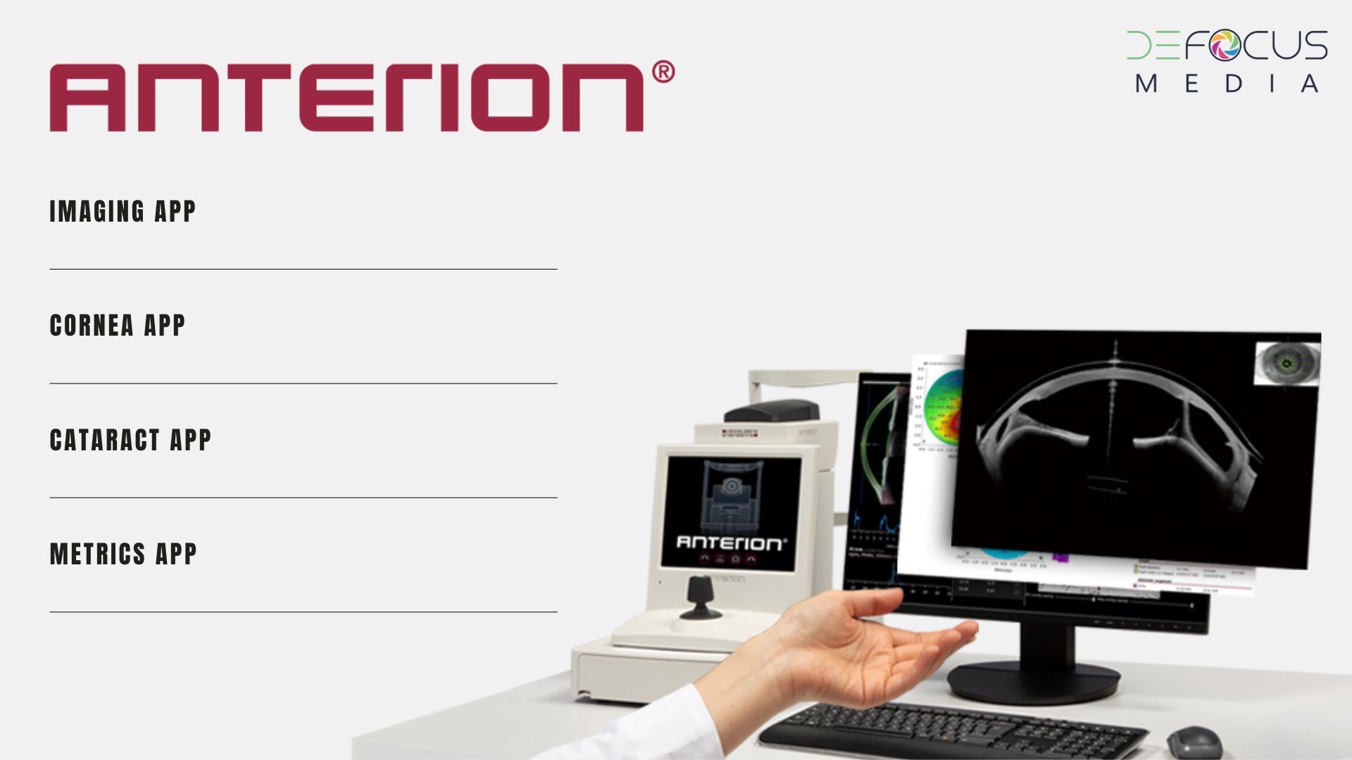
The Role of Heidelberg Engineering Anterion in Clinical Decision-Making
This is where advanced imaging became essential. The Heidelberg Engineering Anterion provides wide-field anterior segment OCT imaging, allowing clinicians to visualize up to 16.5 mm across the anterior segment. This capability enables a comprehensive assessment of:
- Full scleral lens vault from limbus to limbus
- Clearance over the graft-host junction
- Localized pachymetry changes beneath the lens
- Lens geometry relative to oblate or prolate corneal shapes
In post-PKP eyes, a lens may appear ideal centrally while exerting subtle stress in the mid-periphery. Without wide-field imaging, these issues can go undetected until corneal health begins to decline.
Baseline Imaging as a Preventive Strategy
One of the strongest clinical takeaways from the discussion was the importance of imaging asymptomatic patients. Establishing baseline anterior segment OCT data allows clinicians to:
- Compare future scans when symptoms arise
- Distinguish true progression from longstanding anatomy
- Avoid unnecessary scleral lens redesigns
- Detect early hypoxia-related edema before vision is affected
Rather than waiting for visual decline, proactive imaging supports earlier, more confident intervention—often preserving both corneal health and lens success.
Medication Adherence: The Quiet Threat to Graft Survival
Once imaging confirmed that the scleral lens was not the primary issue, attention turned to medication adherence. Similar to glaucoma management, graft rejection can progress silently until vision is compromised.
The panel emphasized that re-education must be ongoing, particularly as follow-up intervals extend. Patients may interpret clinical stability as permission to discontinue therapy, making it essential to reinforce:
- The purpose of long-term steroid use
- The risks associated with discontinuation
- Practical strategies to improve adherence
In this case, restoring compliance was far more impactful than altering lens parameters.
Replacing a Stable Scleral Lens: Clinical Guidance
When a scleral lens has performed well over time, the recommended approach is conservative:
- Begin with a duplicate lens design
- Avoid chasing small over-refractions caused by lens warpage
- Make incremental improvements over time rather than dramatic changes
Clinical stability and corneal health, not the pursuit of a “perfect” image, should guide lens replacement decisions.
This Grand Rounds LIVE case reinforces that modern scleral lens care extends well beyond achieving good visual acuity. By integrating patient education, thoughtful lens management, and advanced imaging with tools such as the Heidelberg Engineering Anterion, eye care professionals can better protect corneal health, extend graft longevity, and improve long-term outcomes for complex patients.
Ultimately, the most meaningful clinical decisions often come not from changing the lens—but from understanding the eye beneath it.